Dilema Clinico
Paciente Belga, 38 años, solo habla neerlandés , esposa Dominicana, hace tres días llega al país, ya viene con malestares, fiebre, desorientación, estado confusional, alucinaciones, ictericia ( color amarillo de piel y conjuntivas),equimosis ( sangrados difusos por debajo de la piel ),historia de stress severo y de fuerte alcoholismo ( de años). Analítica: Bilirrubina elevada ( las 2 ,directa e indirecta ), alteración de pruebas hepáticas, plaquetas bajas y descendiendo en análisis sucesivos. Pruebas del Dengue negativas. La tomografía Axial Computarizada ( TAC) muestra aparente nódulo en lóbulo cuadrado, se realizó otra con contraste, que no aportó nada significativo.
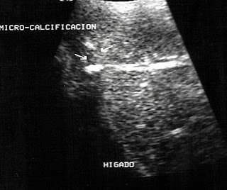
La sonografía que le realicé , mostró hepato-esplenomegalia y un lóbulo cuadrado aumentado de tamaño e hipo-ecogenico-ver foto-
El paciente fue ingresado en la medianoche del sábado a Domingo, en la mañana del Domingo estábamos discutiendo su caso y actuando : un gastroenterólogo, un Neurólogo, un infectólogo y yo como sonografista.
Se ordenó su traslado a UCI ( con la oposición de la esposa), por el estado confusional ( se había retirado el catéter venoso en 7 ocasiones ) y por el peligro del sangrado por la plaquetopenia ( plaquetas bajas ).Se están barajando los diagnósticos de Síndrome de Abstinencia , Rickettsia ( el dengue y la Hepatitis A,B y C ya fueron descartadas ) y se está a la espera de la analíticas complementarias
Clinical Dilemma
Belgian patient, 38 years old, only speaks Dutch, Dominican wife, three days ago he arrived in the country, he already comes with discomfort, fever, disorientation, confusional state, hallucinations, jaundice (yellow skin and conjunctiva), ecchymosis (diffuse bleeding underneath the skin), history of severe stress and strong alcoholism (years). Analytical: High bilirubin (2 o'clock, direct and indirect), alteration of liver tests, low platelets, and decreasing in successive analyzes. Dengue tests negative. The Computed Axial Tomography (CT) shows an apparent nodule in a square lobe, another with contrast was performed, which did not provide anything significant.
The sonography I performed showed hepato-splenomegaly and an enlarged and hypo-echogenic square lobe-see photo-
The patient was admitted at midnight from Saturday to Sunday, on Sunday morning we were discussing his case and acting: a gastroenterologist, a neurologist, an infectious disease specialist, and myself as a sonographer.
His transfer to the ICU was ordered (with the opposition of the wife), due to the confusional state (the venous catheter had been removed on 7 occasions) and the danger of bleeding due to plateletpenia (low platelets). Withdrawal Syndrome, Rickettsia (dengue and Hepatitis A, B and C have already been ruled out) and complementary tests are being awaited


