El Poder de la Sonografia
Poder hacer un diagnostico en localizaciones corporales difíciles es una de las virtudes de la sonografía como método diagnostico. Este caso me recordó otro similar durante el cual el urólogo tratante me afirmaba que en esa localización ( uréter bajo ) era imposible localizar un calculo de pequeñas dimensiones. Yo, le miré sorprendido y en tono retador, le dije: Pues yo siempre lo he logrado ! , No suele ser ese mi actitud en estos casos, pero me sentí algo ofendido y eso significó un reto para mí. En aquel caso como en éste logré definir la presencia de la pequeña imagen del cálculo, ante lo cual él no tuvo mas remedio que pedirme excusas por haber puesto en duda mi capacidad.
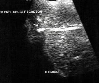
En esta entrega se trata de un pequeño calculo localizado en porción distal del uréter derecho, la paciente femenina de 42 años se presentó con un aparatoso cuadro de intenso dolor en Fosa Iliaca Derecha, nauseas, vómitos. Nótese la sombra posterior que produce el calculo al interrumpir el flujo de sonido que choca contra él. Se descartó durante este mismo examen la presencia de lesiones de ovario derecho o del anexo pelvico homolateral, lo cual demuestra otra de las capacidades de la sonografía para hacer diagnósticos diferenciales " in situ " durante un solo rastreo del área sospechosaThe Power of Sonography
Being able to make a diagnosis in difficult body locations is one of the virtues of sonography as a diagnostic method. This case reminded me of a similar one during which the treating urologist told me that in this location (lower ureter) it was impossible to locate a small stone. I looked at him in surprise and in a challenging tone, I said: Well, I've always done it! This is not usually my attitude in these cases, but I felt somewhat offended and that meant a challenge for me. In that case, as in this one, I managed to define the presence of the small image of the calculus, before which he had no choice but to apologize for having questioned my ability.
In this issue, it is a small calculus located in the distal portion of the right ureter. The 42-year-old female patient presented with a spectacular picture of intense pain in the Right Iliac Fossa, nausea, vomiting. Note the posterior shadow that the calculation produces when interrupting the flow of sound that collides with it. During this same examination, the presence of lesions of the right ovary or of the ipsilateral pelvic annex was ruled out, which demonstrates another of the capabilities of sonography to make differential diagnoses "in situ" during a single scan of the suspicious area.


