Masa Solida Anexo Derecho
Paciente femenina 54 años edad con dolor en Fosa Ilíaca Derecha que se irradia hacia fosa renal homolateral y hacia el muslo hasta la rodilla.
Gesta 2 Partos 2. Histerectomia total & Ooforectomia ( no sabe de cual lado ) a los 48 años de edad.
Hace 2 años sonografia pelvica que detecto masa solida. Tomografía Axial Computarizada ( TAC) que detecta masa solida de unos 4 cm del lado derecho de pelvis-
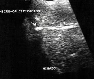
La sonografia abdominal (abdomen superior ) es normal. En la sonografia pelvica transvaginal y transabdominal ( al través de vejiga llena ) se aprecia masa solida redondeada, de limites regulares, localizada en anexo derecho, no aumento significativo del flujo vascular al Doppler Color, mide aprox 4.6 x 4.6 x 3.2 cm.
El nuevo examen del TAC muestra hallazgos similares- ver ultimas imágenes-
Solid Mass Right Annex


Imágenes de la masa en la Tomografía Axial Computarizada (TAC)


