Quiste Mediastino
Paciente femenina de 50 años de edad con antecedente remoto ( 6 años ) de tumoración cerebral operada ( tumor de nervio óptico izquierdo ) con subsecuente perdida de visión de ese ojo, ella dice que tenia 35 años con ese tumor. Gran fumadora. Se queja de insomnio y anorexia. Dolor torácico retro-esternal , punzante, con disnea, fiebre y perdida de peso ( 10- 15 libras ) de tres meses de evolución. Presenta ortopnea, tos persistente, no productiva.
La ecosonografia cardíaca mostró hemo pericardio Hemoglobina Glicosilada de 5,7 ( normal 4-6 )
Glicemia Basal 107
Creatinina 0,6
Examen de orina Normal
Leucocitos 11,700
Hematocrito 37,4
Troponina 0,01
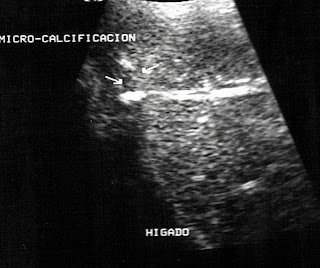
En imágenes vemos las dos primeras con nuestros hallazgos en la sonografía mostrando el quiste mediastínico visto en proyección desde el abdomen de la paciente con tamaño de 17,4 X 12,9 X 8,9 cm.
Las dos imágenes siguientes son de cortes tomográficos de la misma lesión y las ultimas imágenes son angio-tomografias 3 D tórax-pulmón del mismo caso, los cuales son descritos como gran colección de densidad liquida ,compleja, de aprox 16 UH que aparenta pertenecer a pleura izquierda, mide 16 X 15 cm en sentido AP y causa desplazamiento contra-lateral de las estructuras mediastínicas y atelectasias pasivas en pulmón izquierdo. Se aprecia pequeño derrame pleural izquierdo, Aorta Torácica normal y pequeñas adenomegalias inespecíficas.
Mediastinal Cyst
Female patient aged 50 with remote history (6 years) of operated brain tumor (tumor of the left optic nerve) with subsequent loss of vision in that eye, she says she's had 35 years with tumor. Heavy smoker. She complains of insomnia and anorexia. Retro-sternal chest pain , stabbing, with dyspnea, fever and weight loss (10 to 15 pounds) of three months. Presents orthopnea, persistent cough, nonproductive.
Ecosonography the heart showed hemopericardium
Glicosolada hemoglobin with of 5.7 (normal 4-6)
Basal glycemia 107
Creatinine 0.6
Normal Urine
Leukocytes 11.700
Hematocrit 37.4
Troponin 0.01
In the images we see the first two with our findings on sonography showing the mediastinal cyst seen in projection from the abdomen of the patient with size of 17.4 X 12.9 X 8.9 cms.
The following are two images of tomographic slices of the same lesion and the latest images are 3-D tomograms angio-chest-lung of the same case, which are described as a large collection of liquid density, complex of about 16 UH apparently from left pleura, measuring 16 X 15 cm in AP direction and causes contralateral shift of mediastinal structures and left lung atelectasis. Small left pleural effusion is seen with normal thoracic aorta and small nonspecific lymphadenopathy.


Comentarios