Miomas & Quiste Hemorragico & Infertilidad Primaria & Coleccion Liquida
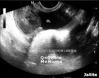
Paciente femenina 29 años de edad, Gesta 0 Para 0, Menarquia los 11 años, Primera Relación Sexual a los 16 años, con infertilidad primaria de 13 años, con cuatro ( 4 ) parejas sexuales. Sufrió Miomectomia de seis ( 6 ) miomas y tratamiento medico para su infertilidad. Se queja de dispareunia, dolores y calambres en Fosa Ilíaca Derecha que se irradian hacia el muslo homolateral. Tiene periodos de Amenorreas. Al examen sonografico vía transvaginal apreciamos útero con múltiples ( 3 ) imágenes nodulares solidas, miomatosas. El mayor se localiza en cuerno izquierdo, mide aprox: 4,2 x 3,3 cm, es sub-seroso, otro mioma, intra- mural, se localiza en cuerpo-fundus, mide aprox: 3,8 x 3,4 cm, el mas pequeño de los tres es intramural, se localiza en cara anterior de cuerpo uterino, mide aprox: 2,3 x 1,3 cm. El ovario derecho muestra imagen anecogena, quistica, con material fibrinoso en forma de bandas, paredes gruesas y refuerzo ecogenico posterior, mide aprox: 4,2 x 3,6 cm. Adicionalmente se visualiza colección liquida con grumos finos en suspensión en las cercanías del quiste ovárico, esto ultimo sugiere hiper-estimulación ovárica medicamentosa.
Fibroids & Primary Infertility & Hemorrhagic Cyst & Liquid Collection
A female patient aged 29, Gesta 0 Para 0, Menarche age 11, first sexual intercourse at age 16 with primary infertility of 13 years, with four (4) sexual partners. Myomectomy suffered for six (6) fibroids and medical treatment for fibroids and infertility. She complains of dyspareunia, pain, and cramps in right lower quadrant that radiates into the thighs homolateral. She has periods of amenorrheas. Transvaginal sonographic examination appreciates uterus with multiple (3) solid nodular fibroids. The greater is located on the left horn, measures approx: 4.2 x 3.3 cms, is sub-serous, another fibroid, intramural, is located in the body-fundus, measures approx: 3.8 x 3.4 cms, the smallest, or all three are intramural, is located in front of the uterine body measures approx: 2.3 x 1.3 cms. The right ovary shows an image-echogenic cystic, with fibrinous material in strip form, thick walls and reinforcing posterior echogenic, measures approx: 4.2 x 3.6 cms. Additionally, the fluid collection is displayed with fine clumps suspended in the vicinity of the ovarian cyst, the latter suggests ovarian hyper-stimulation drug.


