Puede Diagnosticarse La Neumonia por Sonografia ?
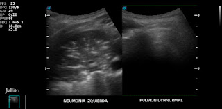
Ponemos este título porque aún hay escépticos o peor aún , colegas desactualizados que no conocen el potencial diagnóstico de la sonografia en las patologías pulmonares. Ponemos un ejemplo de un caso reciente para ilustrar el tema. Recibimos niño de 02 años de edad, lloroso y quejumbroso, con aspecto general de estar realmente enfermo. Los familiares me explican que tiene cuatro ( 4 ) dias de evolucion con fiebres altas, malestar general, inapetencia y tos persistente. La sonografia abdominal nos muestra el aspecto característico de hígado estrellado ( starry sky liver ), al preguntar, los familiares niegan haberle suministrado Acetaminofen, que en nuestro medio es la causa más frecuente que solemos encontrar asociado a estos hallazgos. La vesícula muestra engrosamiento uniforme de sus paredes, compatible con el diagnóstico de colecistitis aguda alitiásica, el hígado se aprecia aumentado de tamaño. Decidimos entonces explorar los pulmones encontrando signos clarísimos de neumonia izquierda, con múltiples focos hiperecogénicos conformando el llamado broncograma aéreo.En la misma imagen ,incluimos el aspecto sonografico del pulmon derecho, el cual es totalmente normal.Posteriormente, hacemos la correlación con la imagen de la radiografía pulmonar tomada un poco antes del examen sonografico y que no habíamos visto hasta entonces. Con toda sinceridad, con pasion, para mi, la imagen sonografica es mas demostrativa de la neumonia, que la radiografia de torax , juzguen ustedes........
It Can Be Diagnosed By Sonography Pneumonia?
We put this title because there are still skeptical or even worse, outdated colleagues who do not know the diagnostic potential of sonography in lung diseases. We provide an example of a recent case to illustrate the issue. We receive child 02 years old, tearful and plaintive, the general appearance of being really sick. Relatives tell me that it has four (4) days of evolution with high fever, malaise, loss of appetite and a persistent cough. Abdominal sonography shows the characteristic appearance of the starry liver (liver starry sky), asking, family deny having supplied acetaminophen, which in our area is the most frequent cause we usually find associated with these findings. The gallbladder shows uniform thickening of its walls, consistent with the diagnosis of acute cholecystitis acalculous, liver shown enlarged. Then we decided to explore the lungs finding very clear signs of pneumonia left, forming the reflective foci called air bronchograms. In the same image, we include the sonographic appearance of the right lung, which is totally normal. Later, we make the correlation with the image of lung x-ray taken shortly before the sonographic examination and we had not seen before. In all honesty, with passion, for me, the sonographic image is more demonstrative of pneumonia, the chest x-ray, you judge ........


