Aneurisma Aortico
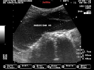
Paciente femenina , 88 años de edad. Hipertensa, delgada, fumadora inveterada de pipa. Toma muchas tazas de café a diario. A la exploración física se aprecia tumoración abdominal pulsatil. Al examen sonografico abdominal se visualiza dilatación aneurismatica aortica de 11.7 X 4,3 X 3.4 cm, localizado por debajo de la salida de la arteria renal izquierda. El aneurisma muestra pared engrosada por trombos fijos adosados a ella ( grosor 1.4 cm ). En todo el trayecto examinado se aprecian múltiples placas ateromatosas. La Vena Cava inferior también luce muy aumentada de calibre en su trayecto intrahepatico.
Female patient, 88 years old. Hypertensive, slim, inveterate pipe smoker. It takes many cups of coffee daily. A physical examination can be seen pulsating abdominal tumor. Sonographic examination abdominal aortic aneurysmal dilation is displayed 11.7 X 4.3 X 3.4 cm, located below the outlet of the left renal artery. The thickened wall shows an aneurysm by thrombus attached to its fixed thickness (1.4 cm). All the way discussed multiple atheromatous plaques are seen. The inferior cave vein also increased in size looks great in its way intrahepatic
Aortic Aneurysm
Female patient, 88 years old. Hypertensive, slim, inveterate pipe smoker. It takes many cups of coffee daily. A physical examination can be seen pulsating abdominal tumor. Sonographic examination abdominal aortic aneurysmal dilation is displayed 11.7 X 4.3 X 3.4 cm, located below the outlet of the left renal artery. The thickened wall shows an aneurysm by thrombus attached to its fixed thickness (1.4 cm). All the way discussed multiple atheromatous plaques are seen. The inferior cave vein also increased in size looks great in its way intrahepatic


