Tumor Testicular
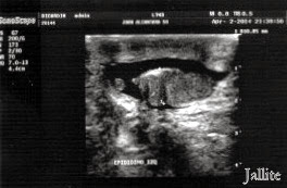
Paciente de 58 años de edad que consulta por impotencia relativa relacionada al parecer por un medicamento anti-hipertensión que lleva usando desde hace unos meses. Anteriormente , con otro anti-hipertensivo, no le sucedía. Durante el examen , refirió que sufre de molestias dolorosas en los testículos, sobre todo en el derecho, por tal motivo llega a nosotros referido para examen sonografico testicular. El testículo derecho muestra heterogeneidad focal a expensas de dos áreas hipoecogenicas, de fronteras irregulares y de difícil delimitación . Ambos epidídimos lucen engrosados, edematosos, con presencia de dos quistes en el derecho y de tres ( 3 ) quistes en el izquierdo. El testículo izquierdo muestra presencia de múltiples imágenes quisticas simples, las mayores miden aprox : 14,5 X 10,1 mm, 5,3 X 3,5 mm y 3,8 X 3,0 mm respectivamente. Se aprecia ademas hidrocele con grumos finos sedimentarios en el testículo izquierdo. Se concluye con los siguientes diagnósticos :
1.-Probable Tumoración Sólida Testiculo Derecho
2.-Epididimitis Bilateral
3.-Quistes Testiculo Izquierdo
4.-Quistes Epididimos ( Bilateral )
5.-Hidrocele Izquierdo
Con este caso se demuestra la gran capacidad de la eco-sonografia de detectar lesiones testiculares diferentes en su origen, evolución y dar la posibilidad de actuar sobre diversos aspectos lesionales en un mismo paciente.
Pensamos que el diagnostico mas probable es el de Seminoma Multifocal de testículo derecho pero hay que hacer diagnóstico diferencial con otros tipos tumorales y posiblemente sea la biopsia testicular la que diga la última palabra.
Testicular Tumor
A
58 -year-old relative impotence clinics related apparently by an anti
-hypertension drug that has been using for a few months. Previously, with other antihypertensive, not happened. During the discussion, he said that he suffers from painful discomfort in the testicles, especially on the right, as such comes to us referred for testicular sonographic examination. The right testis shows focal heterogeneity at the expense of two hypo -
echogenic, irregular border areas, and difficult delimitation. Both look thickened epididymis, edematous, with the presence of two cysts on the right and three (3 ) cysts on the left. The left testicle shows the simple cystic presence of multiple images, elderly measure approx: 14.5 X 10.1 mm, 5.3 X 3.8 mm, and 3.5 mm 3.0 mm respectively. A hydrocele is seen also with fine sediment lumps in the left testicle. It concludes with the following diagnoses :
1.- Probable Solid tumor Right Testicle
2.- Bilateral epididymitis
3.- Left Testicular Cysts
4.- Epididymal cysts ( Bilateral)
5.- Left Hydrocele
This case demonstrates the great capacity of the eco- sonography to detect different testicular lesions in their origin, evolution and give the possibility to act on various aspects of a lesion in the same patient.
We think the most likely diagnosis is that of multifocal seminoma of right testicle but to do differential diagnosis with other tumor types and possibly testicular biopsy which says the last word.
Ref Malignant Testicular Tumor Imaging
Testicular Ultrasound & Pathology of the Testes
Sonography of the Scrotum
 |
| Epididimitis Izquierda |
 |
| Quistes Testículo Izquierdo |
 |
| Quistes Epididimo Izquierdo |
 |
| Quistes Epididimo Izquierdo & Hidatides |
 |
| Hidrocele Izquierdo |
 |
| Tumoración Testículo Derecho |
 |
| Tumoración Testículo Derecho |
 |
| Tumoración Testículo Derecho |
 |
| Tumoración Testículo Derecho |


