Hemangioma Hepatico Diagnosticado Intrautero
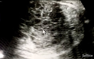
Paciente de 25 años de edad cursando con su segundo embarazo. Fue evaluada por otro sonografista hace 15 dias reportándose ¨imagen eco mixta abdominal para evaluar después del nacimiento del producto¨.El medico referidor quiso repetir el examen a los 15 dias para un diagnostico mas acabado.En nuestro examen encontramos embarazo de 37,0 semanas, cefálico,longitudinal, con peso estimado de 3,631 gramos ( 7,9 libras ). Muestra corazón, ambos riñones, vejiga, columna vertebral,cráneo y extremidades dentro de la normalidad. A nivel abdominal se aprecia masa con aspecto esponjoso que ocupa gran parte del abdomen fetal, aumentando la circunferencia abdominal, la masa mide aprox: 8,60 X 7,07 cm. Es ecoDoppler negativa. Se visualiza polihidramnios leve.Se concluye con el diagnostico de Quiste hepático y Polihidramnios leve. El producto, femenino, nace por cesárea con peso estimado similar al calculado previamente, se evalúa a las 2 horas de nacer por sonografia la cual confirma los hallazgos previos al nacimiento. Se concluye con el diagnostico de Hemangioma hepático de carácter esponjoso y de común acuerdo con el cirujano pediátrico, se acuerda manejo conservador con vigilancia del proceso.
Intrauterine Diagnosed Hemangioma
Patient of 25 years of age studying with her second pregnancy. It was evaluated by another sonographer 15 days ago reporting "abdominal echo image to evaluate after the birth of the product". The referral doctor wanted to repeat the test at 15 days for a more finished diagnosis. In our examination, we found a pregnancy of 37.0 weeks, cephalic, longitudinal, with an estimated weight of 3.631 grams (7.9 pounds). Shows heart, both kidneys, bladder, spine, skull, and extremities within normal. At the abdominal level, a spongy mass can be seen that occupies a large part of the fetal abdomen, increasing the abdominal circumference, the mass measures approx: 8.60 X 7.07 cm. It is negative echo doppler. Mild polyhydramnios is visualized. It concludes with the diagnosis of a hepatic cyst and mild polyhydramnios.
The product, female, is born by cesarean section with an estimated weight similar to that previously calculated, it is evaluated 2 hours after birth by sonography, which confirms the findings prior to birth. It concludes with the diagnosis of hepatic hemangioma of spongy character and in agreement with the pediatric surgeon, conservative management is agreed with monitoring of the process.
The product, female, is born by cesarean section with an estimated weight similar to that previously calculated, it is evaluated 2 hours after birth by sonography, which confirms the findings prior to birth. It concludes with the diagnosis of hepatic hemangioma of spongy character and in agreement with the pediatric surgeon, conservative management is agreed with monitoring of the process.
 |
| Imagen del primer examen ( fuente externa) Image of the first exam (external source) |
 |
| Imagen del primer examen ( fuente externa) Image of the first exam (external source) |
 |
| Imagen del primer examen ( fuente externa) Image of the first exam (external source) |
 |
| Imagen del riñón derecho normal Image of the normal right kidney |
 |
| Doppler de la masa quistica Doppler of the cystic mass |
 |
| Doppler de la masa quistica Doppler of the cystic mass |
 |
| Doppler de la masa quistica Doppler of the cystic mass |
 |
| masa quistica cystic mass |
 |
| Doppler Área Cardíaca y Aorta Abdominal Doppler Cardiac Area and Abdominal Aorta |
 |
| Imagen axial del Producto-Tórax-Hígado y Masa Quistica Axial Image of the Product-Thorax-Liver and Cystic Mass |
 |
| Doppler Área Cardíaca Doppler Cardiac Area |
 |
| Imagen Hígado a las 2 horas del Nacimiento Liver image at 2 hours of birth |
 |
| Riñon Derecho a las 2 horas del Nacimiento Kidney Right at 2 o'clock in Birth |
 |
| Doppler del Hemangioma a las 2 horas de nacer Doppler of the Hemangioma at 2 hours of birth |
 |
| Vesicula a las 2 horas de nacer Gallbladder at 2 hours of birth |
 |
| Hemangioma Hepatico a las 2 horas de Nacer Hepatic Hemangioma at 2 hours of birth |
 |
| Hemangioma Hepatico a las 2 horas de Nacer Hepatic Hemangioma at 2 hours of birth |


