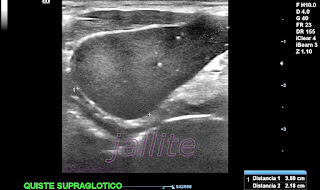
Quiste Branquial Supraglotico
Masculino 68 años de edad, profesor universitario, sin antecedentes personales de interés que presenta desde hace 1 año ligera disfonía y cierto grado de ronquera con disfagia leve. El examen físico muestra nódulo duro, móvil, localizado inmediatamente por debajo del área de la glándula submaxilar derecha. La palpación no es dolorosa. Se le realiza tomografía axial computarizada la cual muestra tumoración supraglótica que comprime/desplaza la tráquea (fotos TAC #1 y2). La exploración sonográfica demostró una imagen quística con grumos que ocupan toda la luz y con presencia de punteado hiperecogénico (microcalcificaciones ) disperso. Las paredes son finas y hay pobre refuerzo ecogénico posterior debido a la densidad del contenido quístico. La masa quística mide aprox: 1.69 X 2.18 X 2.17 cm con un volumen aprox: de 9.15 c.c.Una muestra fue cultivada para detectar proceso infeccioso y fue reportada como negativa.El tejido extraido fue reportado por el patologo como: estructura quistica benigna tapizada predominantemente por epitelio escamoso estratificado sin atipia asociada a inflamacion cronica activa focal y areas de necrosis. Diagnostico diferencial entre quiste laringeo ductal /sacular y laringocele entre otros.
Este caso es muy similar a otro publicado por nosotros en Julio del 2022-Quiste Branquial Recidivante de Cuello
Supraglottic Branchial Cyst
A 68-year-old male, a university professor, with no personal history of interest has presented mild dysphonia and some degree of hoarseness with mild dysphagia for the past 1 year. Physical examination shows a rugged, mobile nodule below the right submandibular gland area. Palpation is not painful. A computerized axial tomography was performed, which showed a supraglottic tumor that compressed/displaced the trachea (photos TAC 1-2). Sonographic examination revealed a cystic image with lumps occupying the entire lumen and the presence of scattered hyperechogenic (microcalcifications) stippling. The walls are thin and there is poor posterior echogenic enhancement due to the density of the cystic contents. The cystic mass measures approximately 1.69 X 2.18 X 2.17 cm with a volume of approximately 9.15 c.c.One sample was cultured to detect an infectious process and was reported as negative. The extracted tissue was reported by the pathologist as a benign cystic structure lined predominantly by squamous stratified epithelium without atypia associated with focal active chronic inflammation and areas of necrosis. Differential diagnosis between ductal/saccular laryngeal cyst and laryngocele among others.
This case is very similar to another published by us in July 2022-Recurrent Branchial Cyst of the Neck.
 |
| #3 |
 |
| TAC #1 |
 |
| TAC #2 |
 |
Tras cirugía con enucleación del quiste se crea nuevo nódulo en el área quirúrgica, el examen sonografico muestra imagen anecogena, quística, mixta (liquido con material ecogeno trabeculado en su interior). Las paredes son finas y se aprecia refuerzo ecogénico posterior y muestra refuerzo ecogénico posterior. Mide aprox: 2.09 X 1.45 X 1.43 cm con un volumen aprox: de 2.63 c.c. Se concluye con el diagnostico de colección hemática residual.
After surgery with enucleation of the cyst, a new nodule is created in the surgical area, and the sonographic examination shows an anechoic, cystic, mixed image (liquid with trabeculated echogenic material inside). The walls are thin and posterior echogenic enhancement is appreciated and shows posterior echogenic enhancement. It measures approx: 2.09 X 1.45 X 1.43 cm with a volume of approx: 2.63 c.c. It concludes with the diagnosis of residual blood collection.
 |
| #1 -Imagen Post-Cirugia |
 |
| #2 Imagen Post-Cirugia |
 |
| #3 Elastografia Quiste Residual Post-Cirugia |


Comentarios