Calculo Conducto Biliar Secundario
Los casos complejos llevan una evolución cuyo final es difícil de predecir.Mostramos este caso como ejemplo de esta aseveración. Se trata de paciente masculino de 60 años de edad que inicialmente vimos ,de emergencia, a altas horas de la noche, ingresado en cuidados intensivos, en coma, intubado, etc,. Eso fue el 20 de Abril 2009. El paciente tenía como antecedente inmediato una cirugía de Bypass Aórtico abdominal, realizada un mes antes de su ingreso, el examen abdominal mostró como único hallazgo una Colecistitis Aguda A-litiasica, edematosa, con una pared anterior de 14 mm ( normal hasta 3 mm ) y derrame liquido peri-vesicular, el contenido vesicular de bilis estaba reducido debido al edema parietal , se midió 8.98 c.c.
Tres días mas tarde, un nuevo examen mostró que el paciente, comatoso aún, tenia su colecistitis aguda A-litiasica con una reducción del grosor de la pared anterior hasta 10 mm en lugar de los 14 mm pero ademas, se había agregado leve derrame pleural derecho , aumento del calibre de la vena esplénica y esplenomegalia de 489 c.c., junto a signos incipientes de insuficiencia renal crónica ( IRC ).
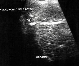
Ya para el 08 de mayo, se apreció la presencia de calculo localizado en vía biliar secundaria, rama izquierda, con presencia de sombra ecogenica posterior-ver fotos-El derrame pleural derecho ha desaparecido y la vesícula muestra una pared anterior fina de unos 3 mm ( normal ) con un contenido biliar claro y un volumen de 38.0 c.c. Junto a esto esplenomegalia , ya reducida de 489 c.c. a 433 c.c.
El paciente se ha recuperado del coma,esta en una habitación normal, conversa y bromea con todos, solo preocupa su analítica que muestra incremento creciente de la bilirrubina total ( 7.6 mlg/dl con directa de 5.9 dl ) con GOT/AST de 125 u/l ( normal hasta 41 u/l) GPT /ALT de 103 u/l ( normal hasta 42 u/l ) .
Secondary Bile Duct Calculus
Complex cases take an evolution whose end is difficult to predict. We show this case as an example of this. These are male patients aged 60 years who initially saw, emergency, in the middle of the night, admitted to intensive care in a coma, intubated, and so on. That was on April 20, 2009. The patient was an immediate antecedent abdominal aortic bypass surgery, a month before admission, abdominal examination findings showed how a single acute cholecystitis-lithiasis, edematous, with an anterior wall of 14 mm (normal up to 3 mm) stroke and reduced vesicular fluid, the gallbladder bile was due to parietal edema, was measured 8.98 cc
Three days later, a further examination showed that the patient, still comatose, had its A-lithiasis acute cholecystitis with a reduction of wall thickness above 10 mm instead of 14 mm but had also been added mild pleural effusion right, increasing the size of the spleen and splenic vein of 489 ccs, with emerging signs of chronic renal failure (CRF).
Now for the May 08, showed the presence of bile duct calculus located in the secondary left arm, with the presence of echogenic shadow rear-view photos-The right pleural effusion had disappeared and the gallbladder shows a thin anterior wall of about 3 mm (normal) with clear bile and a volume of 38.0 ccs Along with this splenomegaly, already reduced from 489 ccs 4 to 33 d.c.
The patient has recovered from the coma, is in a normal room, chatting and joking with everyone, just worried about their analytical sample of the growing increase in total bilirubin (7.6 MLG / dl with direct of 5.9 dl) with GOT / AST 125 or / l (normal up to 41 u / l), GPT / ALT 103 u / l (normal up to 42 u / l).


