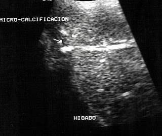
Paciente masculino 86 años de edad ingresa por dolores abdominales difusos. El examen sonografico abdominal muestra hígado aumentado de tamaño con presencia de pequeña imagen nodular, hiper-ecogenica, compatible con el diagnostico de angioma hepático, el hígado muestra aspecto micro-nodular con hipo-ecogenicidad generalizada compatible con hepatopatia crónica descompensada por presentar signos de derrame pleural derecho y ascitis junto a aumento del diámetro de la vena Cava Inferior. En el lóbulo derecho se aprecia pequeña imagen nodular solida , hiper-ecogenica, compatible con el diagnostico de Angioma Hepático. Se aprecia ascitis sub-capsular y peri-vesicular. La vesícula luce casi totalmente ocupada por grumos finos y muestra presencia de múltiples imágenes hiper ecogenicas compatible con colelitiasis múltiple, uno de los cálculos se visualiza en el infundibulo vesicular y otro en conducto cístico, el cual luce agrandado y deformado. Se concluye con el diagnostico de Hepatopatia Crónica Descompensada, Angioma Hepático, Colecistitis con Colelitiasis múltiple y Calculo enclavado en conducto Cístico. Ante una analítica que muestra aumento continuo de la bilirrubina directa, se decide operar, lo cual se realiza a pesar de los riesgos implícitos del caso, todo termina exitosamente y el paciente fue enviado a casa en unos días.
Calculous Cystic Duct
A male patient, 86 years of age admitted for diffuse abdominal pain. The abdominal sonographic examination shows enlarged liver with the presence of the small nodular image, hyper-echogenic, compatible with the diagnosis of hepatic angioma, liver sample micro-nodular hypoechoic with widespread support decompensated chronic liver disease due to signs of pleural effusion right and ascites with the increased diameter of the inferior vena cava. In the right lobe, a small solid, hyperechoic, compatible nodular image with Hepatic Angioma diagnosis is appreciated. Subcapsular and peri-gallbladder ascites are shown. The gallbladder looks almost fully occupied by fine lumps and shows the presence of multiple images hyperechogenic support multiple cholelithiases, one estimate is displayed in the gallbladder infundibulum and one in the cystic duct, which looks enlarged and deformed. It concludes with the diagnosis of chronic liver disease Decompensated, Angioma Hepatic cholelithiasis, and cholecystitis with multiple simulators set in the cystic duct. Given an analytic showing continuous increase in direct bilirubin, it was decided to operate, which is done despite the risks of the case, everything ends successfully and the patient was sent home in a few days.
 |
| Angioma Hepático |
 |
| Grumps en Vesícula |
 |
| Venas Suprahepaticas |
 |
| Angioma & Derrame pleural Derecho |
 |
| Cálculos Vesícula |
 |
| Grumos & Cálculos en Vesícula y Conducto Cístico |


