Historia Natural del Dengue
Imágenes muy demostrativa de la historia natural del Dengue y lo valioso que es el seguimiento de las recomendaciones para valorar la evolución eco-sonografica de esta enfermedad. Recomendamos insistentemente un examen eco-sonografico abdominal al ingreso del paciente, no importando cuantos días lleve desde la aparición de los síntomas y un nuevo examen al 5-6to día de la evolución, este ultimo mostrara la mayor cantidad de datos y permitirá establecer las pautas de seguimiento posterior, por ultimo antes de dar de alta al paciente sugerimos una valoración de egreso, independientemente de como se sienta el paciente.
En este caso el paciente fue examinado a los tres días del comienzo de los síntomas, presentaba fiebre alta, malestar general, dolores retro-oculares, nauseas, vómitos y diarreas, sin embargo, el examen eco-sonografico solo mostró esplenomegalia de 468,9 ml. El resto de los órganos abdominales lucían normales. Al 5to día del comienzo de sus síntomas el paciente se sentía bastante bien desde el punto de vista clínico: no fiebre, no nauseas, vómitos o diarreas y sin cefaleas, sin embargo, el nuevo examen eco-sonografico mostró que la pared anterior de la vesícula tenia un aumento de grosor desde los 4 mm previos hasta 18 mm, con aspecto claramente edematoso, se aprecia ademas derrame pleural bilateral, mas intenso en lado derecho y la esplenomegalia siguió aumentando desde 468,9 ml hasta 516 ml.
El mayor sorprendido fue el paciente al anunciarle que, a pesar de sentirse mejor, la eco-sonografia mostraba signos de posibles complicaciones y que por tanto no podría ser dado de alta todavía.
Very demonstrative images of the natural history of dengue and how valuable it is to follow the recommendations to assess the eco-sonographic evolution of this disease. We strongly recommend an abdominal sonographic examination at patient admission, regardless of how many days lead from the onset of symptoms and a review of 5-6th day of evolution, the latter showed the greatest amount of data and will establish guidelines of follow-up, before finally discharging the patient suggests a review of expenditure, regardless of how the patient feels. In this case, the patient was examined after three days of onset of symptoms, high fever, malaise, retro-ocular pain, nausea, vomiting, and diarrhea, however, eco-sonographic examination showed only splenomegaly 468.9 ml. The rest of the abdominal organs looked normal. On the 5th day of onset of symptoms, the patient felt quite well from the clinical point of view: no fever, no nausea, vomiting, or diarrhea, and without headaches, however, the new eco-sonographic examination showed that the anterior wall gallbladder had an increase in thickness from prior 4 mm to 18 mm, clearly looking edema, bilateral pleural effusion was also noted, more intense on the right side and the spleen continued to increase from 468.9 ml to 516 ml. The biggest surprise was the patient to announce that, despite feeling better, eco-sonography showed signs of possible complications and therefore could not be released yet.
En este caso el paciente fue examinado a los tres días del comienzo de los síntomas, presentaba fiebre alta, malestar general, dolores retro-oculares, nauseas, vómitos y diarreas, sin embargo, el examen eco-sonografico solo mostró esplenomegalia de 468,9 ml. El resto de los órganos abdominales lucían normales. Al 5to día del comienzo de sus síntomas el paciente se sentía bastante bien desde el punto de vista clínico: no fiebre, no nauseas, vómitos o diarreas y sin cefaleas, sin embargo, el nuevo examen eco-sonografico mostró que la pared anterior de la vesícula tenia un aumento de grosor desde los 4 mm previos hasta 18 mm, con aspecto claramente edematoso, se aprecia ademas derrame pleural bilateral, mas intenso en lado derecho y la esplenomegalia siguió aumentando desde 468,9 ml hasta 516 ml.
El mayor sorprendido fue el paciente al anunciarle que, a pesar de sentirse mejor, la eco-sonografia mostraba signos de posibles complicaciones y que por tanto no podría ser dado de alta todavía.
Natural History of Dengue
Very demonstrative images of the natural history of dengue and how valuable it is to follow the recommendations to assess the eco-sonographic evolution of this disease. We strongly recommend an abdominal sonographic examination at patient admission, regardless of how many days lead from the onset of symptoms and a review of 5-6th day of evolution, the latter showed the greatest amount of data and will establish guidelines of follow-up, before finally discharging the patient suggests a review of expenditure, regardless of how the patient feels. In this case, the patient was examined after three days of onset of symptoms, high fever, malaise, retro-ocular pain, nausea, vomiting, and diarrhea, however, eco-sonographic examination showed only splenomegaly 468.9 ml. The rest of the abdominal organs looked normal. On the 5th day of onset of symptoms, the patient felt quite well from the clinical point of view: no fever, no nausea, vomiting, or diarrhea, and without headaches, however, the new eco-sonographic examination showed that the anterior wall gallbladder had an increase in thickness from prior 4 mm to 18 mm, clearly looking edema, bilateral pleural effusion was also noted, more intense on the right side and the spleen continued to increase from 468.9 ml to 516 ml. The biggest surprise was the patient to announce that, despite feeling better, eco-sonography showed signs of possible complications and therefore could not be released yet.

Vesícula a los 3 días del comienzo de los síntomas
Gallbladder at 3 days after onset of symptoms

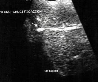
Hígado y Pleura derecha a los tres días del comienzo de los síntomas
Liver and right Pleura on the third day of onset of symptoms

Pared Anterior Vesícula biliar a los 5 días del comienzo de los síntomas
Anterior wall of Gallbladder at 5 days after onset of symptoms

Imagen axial Vesícula Biliar a los 5 días de aparición de los síntomas
Gallbladder axial image at 5 days of onset of symptoms

Derrame Pleural derecho a los 5 días de la aparición de los síntomas
Right Pleural effusion after 5 days of the onset of symptoms

Esplenomegalia a los 5 días aparición síntomas
Splenomegaly at 5 days after onset of symptoms


