Miomatosis Uterina & Ovarios Poliquisticos
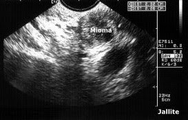
Paciente femenina de 30 años de edad , con historia de infertilidad primaria de 10 años, una sola pareja ( menarquia 14 años, primera relacion sexual a los 20 años ). Llama la atención que hasta los 30 años sus menstruaciones son normales, sin periodos de amenorreas u otros trastornos menstruales. No muestra hirsutismo ni obesidad. Refiere antecedentes familiares de Diabetes Mellitus. Al examen eco-sonografico pelvico vía transvaginal destaca la presencia de tres nódulos miomatosos intramurales, hipo-ecogenicos, localizados en cara postero-lateral derecha de cuerpo uterino ( 3,3 X 3,1 X 3,1 cm ), otro nódulo de características sonograficas similares se localiza en cuerno izquierdo ( 2,3 X 2,2 X 1,9 cm ) y por ultimo el nódulo miomatoso localizado en cara posterior de cuerpo-fundus uterino ( 2,3 X 2,5 X 1,7 cm ). Ambos ovarios lucen aumentados de tamaño con presencia de múltiples quistes de pequeño tamaño ( folículos inmaduros ) , diseminados por todo el parénquima con predominio en zonas periféricas ( ver ultimas dos fotos ). El ovario derecho mide 3,9 X 3,2 X 1,9 cm y el izquierdo 4,8 X 4,1 X 2,7 cm. Se diagnostica pues un cuadro de Miomatosis Uterina y Ovarios Poliquisticos.
A female patient aged 30 with a history of primary infertility of 10 years, one partner (14 years at menarche, first intercourse at age 20.) It is noteworthy that up to 30 years your periods are normal, without periods of amenorrhea or other menstrual disorders. Shows no hirsutism or obesity. Refer a family history of Diabetes Mellitus.
The eco-sonographic examination transvaginal pelvic highlights the presence of three intramural fibroid nodules, hypo-echogenic, located in the posterior-lateral right side of the uterus (3.3 X 3.1 X 3.1 cm), another of similar sonographic characteristic nodules located in the left horn (2.3 X 2.2 X 1.9 cm) and finally the fibroid nodule located in the posterior uterine body-fundus (2.3 X 2.5 X 1.7 cms).
Both ovaries look enlarged with the presence of multiple cysts of small size (immature follicles), scattered throughout the parenchyma, predominantly in peripheral areas (see last two photos). The right ovary measures 3.9 X 3.2 X 1.9 cm and left 4.8 X 4.1 X 2.7 cm.
Diagnosed as a picture of multiple fibroids and polycystic ovaries.
Polycystic Ovaries & Uterine fibroids
A female patient aged 30 with a history of primary infertility of 10 years, one partner (14 years at menarche, first intercourse at age 20.) It is noteworthy that up to 30 years your periods are normal, without periods of amenorrhea or other menstrual disorders. Shows no hirsutism or obesity. Refer a family history of Diabetes Mellitus.
The eco-sonographic examination transvaginal pelvic highlights the presence of three intramural fibroid nodules, hypo-echogenic, located in the posterior-lateral right side of the uterus (3.3 X 3.1 X 3.1 cm), another of similar sonographic characteristic nodules located in the left horn (2.3 X 2.2 X 1.9 cm) and finally the fibroid nodule located in the posterior uterine body-fundus (2.3 X 2.5 X 1.7 cms).
Both ovaries look enlarged with the presence of multiple cysts of small size (immature follicles), scattered throughout the parenchyma, predominantly in peripheral areas (see last two photos). The right ovary measures 3.9 X 3.2 X 1.9 cm and left 4.8 X 4.1 X 2.7 cm.
Diagnosed as a picture of multiple fibroids and polycystic ovaries.


