Polipo Endocervical & Apendicitis
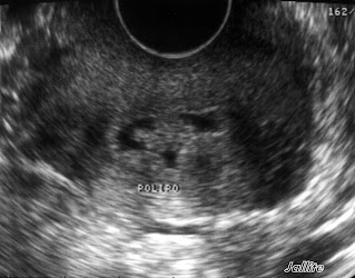
Paciente femenina de 40 años de edad diagnosticada por nosotros de Colelitiasis múltiple hace dos meses, debido a eso fue intervenida. Viene ahora referida por su ginecóloga para sonografia pelvica vía trans-vaginal por proceso febril, nauseas y dolor en fosa iliaca derecha. Aporta un hemograma donde se resalta una leucocitosis de 16,500 con desviación izquierda de la formula. Al examen físico dolor selectivo en el área de la referida fosa con signo de rebote positivo. El examen sonografico muestra un útero aumentado de tamaño ( 10,4 X 5,32 X 5,85 cm ) con presencia de canal cervical dilatado y ocupado parcialmente por masa ecogena, con base de implantación claramente demarcada, bordes irregulares, festoneados. La masa muestra aumento del flujo arterial ( ver foto del Doppler ), mide aprox 1,4 X 1,2 X 0.6 cm.
Debido a sus datos clínicos-analíticos procedemos a explorar fosa iliaca derecha donde se aprecia Apéndice Vermiforme aumentado de tamaño ( 12 mm en sentido AP ), por tanto se refiere al Departamento de Cirugía para evaluación desde allí se ordena su ingreso para Apendicectomia de Emergencia.Se trata pues de una paciente que en un periodo muy corto ( dos meses ) ha tenido que enfrentar una Colecistectomia y tiene de frente una Apendicectomia y una extirpación de un pólipo endocervical.
Endocervical Polyp & Appendicitis
A female patient, 40 years of age diagnosed with multiple Cholelithiasis two months ago, was operated on. Comes now referred by her gynecologist for pelvic sonography via trans-vaginal febrile process, nausea, and pain in the right lower quadrant. Provides a CBC which highlights a leukocytosis of 16,500 with a left shift of the formula. Physical examination selective pain in the area of the said pit with positive rebound tenderness. The sonographic examination shows an enlarged uterus (10.4 x 5.32 x 5.85 cm) with the presence of dilated cervical canal and partly occupied by echogenic mass, based implementation clearly demarcated, irregular borders, scalloped. The mass shows increased blood flow (Doppler pictured), measures approx 1.4 X 1.2 X 0.6 cm.
Because of their clinical data-analytic proceed to explore the right quadrant where the vermiform appendix shows enlarged (12 mm in the AP) therefore refers to the Department of Surgery for evaluation if there is an ordered admission to an emergency appendectomy. it is therefore a patient in a very short period (two months) who has faced a cholecystectomy and appendectomy has front and endocervical polyp removal.
Because of their clinical data-analytic proceed to explore the right quadrant where the vermiform appendix shows enlarged (12 mm in the AP) therefore refers to the Department of Surgery for evaluation if there is an ordered admission to an emergency appendectomy. it is therefore a patient in a very short period (two months) who has faced a cholecystectomy and appendectomy has front and endocervical polyp removal.


Comentarios