Adenocarcinoma Gastrico & Metastasis Hepaticas
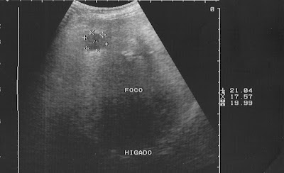
La exploración sonografica mostró: Hígado de tamaño normal con heterogeneidad focal por la presencia de tres (3) áreas hipo-ecogenicas , dos de ellas se muestran como nódulos solidos hipo-ecogenicos de 2.1 X 1.7 X 1.9 cm, otro es de mayor tamaño , pero muestra limites dificiles de precisar, mide aprox: 3.2X 3.8X 3.2 cm y por último una pequeña área quistica de aprox: 1.8X 1.7X 1.4 cm con volumen de 2.06 c.c.*Todas las lesiones se localizan en lóbulo hepático derecho *( ver las dos primeras imagenes ). Hay que hacer notar que hace un mes una tomografía reportó hígado normal, sin lesiones.

The sonographic scan showed normal liver size with heterogeneity focal by three (3) hypo-echogenic areas, two of them appear as hypo-echogenic solid nodules of 2.1 X 1.7 X 1.9 cm, the other is larger, but is difficult to define boundaries, measure approx: 3.2X 3.8X 3.2 cm and finally, a small area of about cystic: 1.8X 1.7X 1.4 cm with a volume of 2.06 cc * All lesions were located in right hepatic lobe * (see The first two images). Note that a month ago, a CT scan reported normal liver without injury.

Las dos próximas imagenes muestran la masa epigastrica,hipo-ecogenica, heterogenea, que mide aprox: 7.1 X 5.5 X 4.3 cm
The next two images show the epigastric mass, hypo-echogenic, heterogeneous, measuring approximately 7.1 X 5.5 X 4.3 cm

History: Female patient 71 years old. Has lost over 20 pounds (9 kilograms) over the past two months. Diabetes mellitus non-insulin-dependent. Hypertension. Presents with bloody vomiting (hematemesis), and dizziness. A physical exam shows mass hard, mobile, not painful in the epigastrium of stony consistency.
The computerized tomography showed mass occupying space in the region of the lesser curvature of the stomach. Normal liver ( a month ago ).
Endoscopy showed gastric mass bulky, friable, easily bleeding, is irregular, located on the first portion of the body, fundus, and lesser gastric curvature.


Comentarios