Quiste Renal
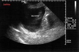
Paciente femenina 42 años de edad, delgada , sin antecedentes patológicos conocidos que desde hace dos meses ha notado masa en hemi-abdomen derecho, móvil, no dolorosa.Niega síntomas urinarios o trastornos intestinales. La exploración eco-sonografica abdominal mostró la presencia de gran masa quistica , no tabicada, que se extiende desde el polo inferior del riñón derecho hasta cresta iliaca homolateral y hasta columna vertebral, desplazando al intestino. Mide 13.1 X 7.6 X 6.9 cm, con un volumen de 360 c.c.
A female patient 42 years old, thin, with no known medical history for the past two months has noticed a mass in right hemiabdomen, mobile, painless. Denies urinary symptoms or intestinal disorders. The eco-sonographic abdominal examination showed the presence of a large cystic mass, not enclosed, which extends from the lower pole of the right kidney and ipsilateral iliac crest up to the spine, displacing the bowel. It measures 13.1 x 7.6 X 6.9 cm, with a volume of 360 ccs.
Renal Cyst
A female patient 42 years old, thin, with no known medical history for the past two months has noticed a mass in right hemiabdomen, mobile, painless. Denies urinary symptoms or intestinal disorders. The eco-sonographic abdominal examination showed the presence of a large cystic mass, not enclosed, which extends from the lower pole of the right kidney and ipsilateral iliac crest up to the spine, displacing the bowel. It measures 13.1 x 7.6 X 6.9 cm, with a volume of 360 ccs.

En la Tomografía Axial Computarizada ( TAC ) con
contraste oral , se apreciaron imágenes confirmatorias
de los hallazgos eco-sonograficos.
In Computed Tomography (CT) with oral contrast,
confirmatory imaging was observed eco-sonographic
findings.
contraste oral , se apreciaron imágenes confirmatorias
de los hallazgos eco-sonograficos.
In Computed Tomography (CT) with oral contrast,
confirmatory imaging was observed eco-sonographic
findings.


