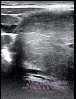
Hipertrofia Nodular Focal del Higado (HNF)
Masculino de 4 años de edad que según la madre ha presentado dese hace 2 semanas un abultamiento en region epigástrica. No se queja de dolor, nauseas, vómitos,fiebre o ningún otro síntoma relevante salvo un ligero proceso diarreico motivo por el cual se le indico coprológico que demostró presencia de amebas y giardias.El examen sonografico abdominal muestra gran masa solida, bien delimitada,que ocupa todo el lóbulo izquierdo del hígado,es isoecogenica con respecto al parénquima hepático vecino,mide aprox: 9.56 X 6.54 cm. Al Doppler color muestra aumento del flujo vascular interno. La elastografia muestra patrón de color con score 2 de Ueno y el estudio con la elastografia shell de una porción de la masa tumoral muestra un patrón compatible con lesión benigna,probablemente una Hiperplasia Nodular Focal (HNF) o un Adenoma Hepatico como 2da posibilidad diagnostica.La hipertrofia nodular focal (HNF) es muy rara ,más frecuente en niños, su frecuencia oscila entre el 0.4 al 0.03 %. Es benigna y su evolucion es indolente.Se recomienda seguimiento.
Ref: 1-Adenoma Hepatico: Caso Clinico
3- Hipertrofia Nodular Focal (HNF)
Focal Nodular Hypertrophy of the Liver (FNH)
A 4-year-old male who, according to his mother, had presented a bulge in the epigastric region for the past 2 weeks. She did not complain of pain, nausea, vomiting, fever, or any other relevant symptom except for a slight diarrheal process, which is why a stool test was indicated, which showed the presence of amoebas and giardia. The abdominal sonographic examination revealed a large, well-defined, solid mass that occupies the entire left lobe of the liver, it is isoechogenic concerning the adjacent liver parenchyma, and it measures approx: 9.56 X 6.54 cm. Color Doppler shows increased internal vascular flow. The elastography shows a color pattern with an Ueno score of 2 and the shell elastography study of a portion of the tumor mass shows a design compatible with a benign lesion, probably Focal Nodular Hyperplasia (FNH) or a Hepatic Adenoma as a 2nd diagnostic possibility. Focal nodular hypertrophy (FNH) is very rare and more frequent in children, its frequency ranges from 0.4 to 0.03%. It is benign and its evolution is indolent. Follow-up is recommended.
Ref: 1-Hepatic Adenoma: Clinical Case
2- Focal Nodular Hyperplasia
3- Focal Nodular Hypertrophy (FNH)
 |
| Nodulo Hepatico con Transductor lineal |
 |
| Nodulo Hepatico Lobulo Izquierdo |
 |
| Lobulo Derecho |


Comentarios