Aneurisma Carotida
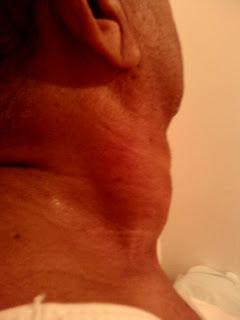
Paciente masculino de 71 años de edad enviado para evaluación de tumoración en lado derecho del cuello ( ver 4 primera fotos ). Antecedentes personales de Diabetes Mellitus tipo II y cardiopatia hipertensiva. El paciente presenta tos no productiva crónica, de unos 25 días de evolución que , según refiere el paciente, coincide en el tiempo con la aparición de la tumoración del cuello.
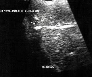
En lado derecho se cuello el examen eco-sonografico muestra arteria carótida con imagen de doble pared ( disecante ? ) ( foto # 5 ) que se comunica con gran masa an-ecogena , con abundantes grumos finos fluctuantes en su interior junto a dos masas ecogenas adheridas a sus paredes. La masa muestra dos segmentos diferenciados, en su parte superior se aprecian múltiples tabiques , esta área mide unos 7,3 x 3,1 cm, la porción mas inferior muestra paredes engrosadas con las masas ecogenas antes mencionadas adheridas a sus paredes, esta área mide aprox: 7,0 x 6,1 x 4,3 cm.( fotos # 6 y siguientes ).
Carotid Aneurysm
Male patient aged 71 referred for evaluation of tumor on the right side of the neck ( see 4 first photos ) Personal history of diabetes mellitus type II a hypertensive heart disease. The patient has a chronic nonproductive cough, about 25 days of evolution, as reported by the patient, coincides with the appearance of the tumor in the neck.
On the right side neck, eco-sonographic examination shows a carotid artery with a double-wall image (dissecting?) (picture # 5) that communicates with great an-echogenic mass, with abundant fine lumps floating in it along with two echogenic masses attached to their walls. The mass shows two distinct segments, at the top, are seen multiple partitions, this area measures about 7.3 x 3.1 cm, the lower portion shows thickened walls with echogenic mass above attached to their walls, this area measures approx: 7.0 x 6.1 x 4.3 cms. (photos # 6 and below).


