Torsión Testicular Crónica con Atrofia Testicular
Masculino 40 años de edad que se presenta con intensos dolores en testiculo izquierdo con irradiaciones hacia la ingle y cara interna del muslo homolateral. Tiene dos meses con las molestias. De vida sedentaria pero practica ejercicios de bicicleta al aire libre, actividad que ha abandonado por el dolor.El examen fisico con palpacion no muestra datos significativos.La sonografia con transductor linear no demuestra la presencia de hernia inguinal izquierda. El examen sonografico testicular muestra un testiculo , epididimo derecho con aspecto sonografico dentro de los parametros normales. El testiculo mide aprox: 3,25 X 2,06 cm. El flujo Doppler es normal.
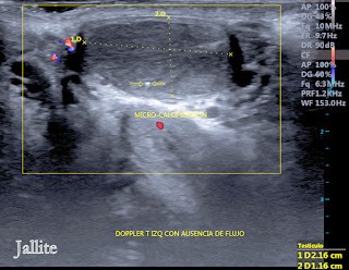
El testiculo izquierdo luce hipoecogenico, de pequeño tamaño, hipotrofico y con total ausencia de flujo vascular al aplicar el Doppler Color. Se visualiza pequeña imagen hiperecogenica, calcificada en su parenquima. El testiculo mide aprox: 2,16 X 1,16 cm.(comparar con el tamaño de su homologo derecho).No hay signos de Varicole con el Doppler Color. Se concluye con el diagnostico de Torsion Testicular Cronica con Atrofia Testicular. El pronostico en estos casos es malo ya que solo queda la opcion de la extirpacion.
Chronic Testicular Torsion with Testicular Atrophy
A 40-year-old male presenting with intense pain in the left testicle with radiation to the groin and inner thigh ipsilateral. He has two months with the discomfort. He was sedentary but practiced bicycle exercises in the open air, an activity that he had abandoned due to pain. Physical examination with palpation did not show significant data. Linear transducer sonography did not show the presence of a left inguinal hernia. The testicular sonographic examination shows a testicle, right epididymis with sonographic appearance within normal parameters. The testicle measures approx: 3.25 X 2.06 cm. Doppler flow is normal.
The left testicle appears hypoechoic, small, hypotrophic, and with a total absence of vascular flow when applying Color Doppler. A small hyperechogenic image is visualized, calcified in its parenchyma. The testicle measures approx: 2.16 X 1.16 cm (compare with the size of its right counterpart). There are no signs of Varicole with Color Doppler. It concludes with the diagnosis of Chronic Testicular Torsion with Testicular Atrophy. The prognosis in these cases is bad since only the option of extirpation remains.


Comentarios