Coleccion Hematica Vaginal
Paciente femenina gesta 3 partos 3, de 34 años de edad que es traída a Emergencias por sangrado transvaginal profuso, agudo. Refiere la paciente que el sangrado comenzó tras mantener unas relaciones sexuales no violentas, según dice, pero con un hombre muy bien dotado. Al examen físico el ginecólogo aprecia amplia herida vaginal que bordea casi 3 /4 parte del cuello uterino, durante el examen físico se constató la presencia de abundante sangre roja, rutilante.
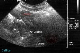
La sonografia pelvica se realiza vía trans abdominal con poca cantidad de orina, para evitar mayores traumas vaginales y previendo que la sangre acumulada impidiera un buen examen. Se pudo apreciar un útero no gestante, en anteversión, cuello uterino normal sonograficamente y la vagina distendida por la presencia de gran colección con abundantes grumos en su interior-ver fotos-.
La herida vaginal fue reparada en quirofano de manera satisfactoria.
Coleccion Vaginal Hematic
Female patient epic 3 part 3, 34 years old who is brought to Emergencies profuse vaginal bleeding, acute. Refer the patient began bleeding after intercourse maintains non-violent, she says, but with a very gifted man. On physical examination, the gynecologist vaginal wound shows wide edge about 3 / 4 of the cervix during a physical examination it was found the presence of abundant red blood, glittering.
The pelvic sonography is performed via trans abdominal with a small amount of urine, to prevent further trauma and vaginal blood collection is anticipating that prevented a good review. We could see a nonpregnant uterus in anteversion, cervix normal and vagina sonographic distended by the presence of a large collection with abundant smooth inside -view photos -.
The vaginal wound was repaired satisfactorily in the operating room.


