Quiste Gigante Tiroides
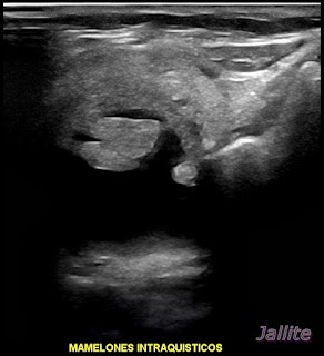
Femenina ,65 años de edad presenta masa visible y palpable en lado derecho del cuello. Según dice, se le presentó repentinamente hace dos meses y ha ido creciendo desde entonces. El examen sonográfico del tiroides con transductor linear de 10.0 MHz muestra gran imagen an-ecogena, quística, contenido claro, con mamelones o excrecencias con crecimiento hacia la luz de la lesión. En estas excrecencias se detecta aumento del flujo al aplicar el Doppler color. Se localiza en el lóbulo derecho, mide aprox: 3.59 X 3.68 X 2.45 cm con volumen aprox: de 16.95 c.c. En ese mismo lóbulo derecho se visualiza otra imagen quística, esta vez con abundantes grumos finos en su interior, mide aprox: 8.7 X 4.6 mm. En lóbulo izquierdo se aprecian dos pequeñas imágenes quísticas de 1.31 X 0.52 cm y de 4.1 X 3.3 mm respectivamente. Para nosotros resulta muy llamativo la rápida evolución ( según la paciente ) del quiste derecho, su gran tamaño y, sobre todo la presencia de excrecencias internas que muestran aumento del flujo al aplicar el Doppler Color todo lo cual nos hace sospechar de lesión maligna.
Giant Thyroid Cyst
Female, 65 years old, presents a visible and palpable mass on the right side of the neck. According to her, she suddenly introduced herself to him two months ago and has been growing ever since. The sonographic examination of the thyroid with a 10.0 MHz linear transducer shows a large an-echogenic, cystic image, clear content, with mamelons or outgrowths with growth towards the lumen of the lesion. In these outgrowths, an increase in flow is detected when applying color Doppler. It is located in the right lobe, measures approx: 3.59 X 3.68 X 2.45 cm with a volume approx: 16.95 c.c. In that same right lobe, another cystic image is visualized, this time with abundant fine lumps inside, measuring approx: 8.7 X 4.6 mm. In the left lobe, two small cystic images measuring 1.31 X 0.52 cm and 4.1 X 3.3 mm, respectively, can be seen. For us, the rapid evolution (according to the patient) of the right cyst, its large size, and, above all, the presence of internal excrescences that show increased flow when applying Color Doppler is very striking, all of which makes us suspect a malignant lesion.


Comentarios